This page is not compatible with Internet Explorer.
For security reasons, we recommend that you use an up-to-date browser, such as Microsoft Edge, Google Chrome, Safari, or Mozilla Firefox.
Scientific Application Examples
Volume Graphics Software in Use
Let Us Help You with Your Research
When scientists require a full-featured, proven, and reliable software for the analysis and visualization of volume data, they choose Volume Graphics software. Whether you’re working in archeology, biology, geology, paleontology or medical research, VGSTUDIO MAX offers features that are perfect for scientific applications.
We're Scientists at Heart
The company Volume Graphics has its roots in science. It was founded in 1997 as a spin-off from a university institute. Back then, Volume Graphics offered the first system for the real-time visualization of computed tomography (CT) data. Today, it’s the solution of choice for thousands of companies – and whenever scientists need to work with volume data.

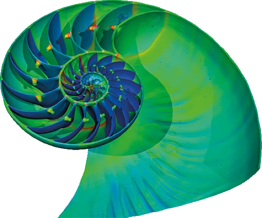
Allonautilus scrobiculatus with color-coded wall thickness

"Incredible 3D renderings, images, and animations"
Benjamin Moreno, Founder and CEO, IMA Solutions
"Incredible 3D renderings, images, and animations"
Benjamin Moreno, Founder and CEO, IMA Solutions"You know each time you use VGSTUDIO MAX that it will work. You will not have any bugs or things like that. And the quality of the rendering is the best in the world. VGSTUDIO MAX enables us to create incredible 3D renderings, images, and animations."
Use the Scanner That Best Fits Your Needs
It doesn’t matter which technique you use for acquiring the image stacks.
Volume Graphics software will work equally well on various kinds of data, such as:
- industrial X-ray CT;
- medical X-ray CT;
- synchrotron tomography;
- neutron tomography; or
- MRI.
Biology
Visualization & Animation
Volume Graphics software makes it easy to create revealing and simultaneously stunning visualizations and animations.* Powerful clipping features allow you to 'look inside' the visualized object without physically dissecting it. And the software can not only produce a photorealistic rendering of any number of volume data sets in one scene, it can also render mesh data sets.
In the case of the brooding South African brittle star Ophioderma wahlbergii**, VGSTUDIO MAX was used to visualize the segmented juveniles inside the mother’s brood pouches. This discovery gave scientists new insights into the brooding behavior of the brittle star.
*Data from J. Landschoff and C. G. Griffiths 2015, ‘3D visualisation of brooding behaviour in two distantly-related brittle stars from South African waters’ and J. Landschoff, A. du Plessis, C. G. Griffiths 2015, ‘A dataset describing brooding in three species of South African brittle stars, comprising seven high-resolution, micro X-ray computed tomography scans’
**Don’t have time to create impressive animations yourself? Let Volume Graphics produce a professional video for you! Contact us for a quote and visit our website and YouTube channel for examples of our work.

Ophioderma wahlbergii, segmented rendering (left) and unsegmented X-ray rendering (right)
Segmentation
In order to define different components, materials, etc., Volume Graphics software offers you powerful yet easy-to-use segmentation tools. The segmentation of fossils or extant organisms within 3D volume data opens up a whole range of possibilities that the conventional thin-sectioning or dissecting of fossils cannot. And all while remaining totally non-destructive. With the available clipping functions, you can cut open a volume object virtually to see what’s inside.
In this example*, the head of the extinct Karusasaurus polyzonus was segmented based on the gray values of the different structures by using the built-in features of VGSTUDIO MAX. Muscle system, bones, and nervous tissue are clearly visible and can be shown and hidden separately.
*Data from E. Stanley and D. Blackburn 2015 (California Academy of Sciences), Scan by: E. Stanley and M. Faillace at GE Inspection Technologies, LP Technical Solutions Center in San Carlos, CA

Karusasaurus polyzonus, segmented muscle system, bones, and nervous tissue
Coordinate Measurement
With VGSTUDIO MAX, you can perform 2D and 3D measurement tasks directly on volume data sets. All you need is the Coordinate Measurement Module. The possibilities go way beyond what can be accomplished when using conventional destructive or other non-destructive examination methods.
On the skeleton of Rhinophrynus dorsalis*, the Mexican burrowing frog, the measurement features were used for anatomical measurements such as the length of extremities or the body. Measurements of anatomical structures like this are used for comparative morphological studies.
*Data from: E. Stanley and D. Blackburn 2015 (California Academy of Sciences), Scan by: E. Stanley and M. Faillace at GE Inspection Technologies, LP Technical Solutions Center in San Carlos, CA

Rhinophrynus dorsalis, measurements of extremities and body length
Advanced Surface Determination*
With the Advanced Surface Determination Module of VGSTUDIO MAX, you can make every detail visible – even those that are smaller than a voxel. Gray values of individual voxels are processed depending on the gray values of the surrounding voxels, giving you a smoother and more realistic surface. This locally adaptive surface determination reduces the influence of artifacts while also minimizing user influence.
The locally adaptive surface determination was used to prepare the skull of the small cat Puma concolor** for highly precise anatomical measurements and accurate visualizations. Precise measurements of anatomical structures can be used for comparative morphological studies.
*Part of the Coordinate Measurement Module
**Data from Volume Graphics GmbH, Scan by: Fraunhofer-Zentrum HTL


Skull of a Puma concolor with a locally adaptive surface determination (white line in 2D slice image)
Thick Slab Option/Non-Planar View
VGSTUDIO MAX can be used to visualize structures that are distributed across several slices of the image stack or to view bent structures. The thick slab option combines consecutive slices into a single 2D view. The non-planar view function 'unrolls' cylindrical objects or flattens a dented surface. What had hitherto only worked with cylindrical objects now also works with freeform surfaces. It enables you to recognize and illustrate bent structures such as inscriptions, adornments, bones or vessels on or within a specimen.
In this example*, the thick slab mode was used to visualize the entire skeleton of a common rat, Rattus norwegicus, in one summarizing 2D view. Originally situated in different slices of the image stack, the inner bone structures became visible at a glance and could therefore be easily followed.
*Data from: Volume Graphics GmbH

Rattus norwegicus, slice image

Thick slab view with complete skeleton
Wall Thickness Analysis
The Wall Thickness Analysis Module for VGSTUDIO MAX can be used to easily determine the wall thickness of objects made of organic or inorganic materials. It helps you to automatically localize and visualize different thicknesses directly within the volume data set.
In this example*, VGSTUDIO MAX was used to examine and visualize the wall thickness of a shell of the marine Allonautilus scrobiculatus, an extant cephalopod. In a 3D rendering, the wall thickness was color-coded, with red indicating thick parts of the shell. The wall thickness analysis can be used to find out more about the adaptation of the species to different water depths.
*Data from: R. Hoffmann 2015 (Ruhr-Universität Bochum), Synchrotron Scan by: F. Fusseis, X. Xiao, R. Hoffmann

Allonautilus scrobiculatus with color-coded wall thickness and related histogram
Structural Mechanics Simulation
With the Structural Mechanics Simulation Module for VGSTUDIO MAX, you can perform virtual stress tests directly on your scanned object. Calculate and visualize force lines, local displacements, and failure-related variables such as von Mises stress.
The Structural Mechanics Simulation Module was used to simulate the bite force on different tooth types of the venomous viper from sub-Saharan Africa (Causus rhombeatus). In the simulation, direct force was applied to the tip of the fang. The measured von Mises stress of a fang was notably lower than when the same force is applied to a standard tooth. This leads to the conclusion that the much larger fangs can withstand a significantly higher load. This type of analysis helps scientists to understand how fang morphology adapts to withstand bite forces, how this differs between fang types, and whether it relates to the feeding behaviors of the respective snakes.
*Data from du Plessis, A., le Roux, S. G., & Broeckhoven, C. (2016), Scan by: Stellenbosch CT Scanner Facility


Causus rhombeatus , visualized force lines showing the simulated bite force
Archaeology
Fiber Composite Material Analysis
The Fiber Composite Material Analysis Module for VGSTUDIO MAX enables you to process both small- and large-scale volume data sets of fiber materials. In small-dimension material samples, VGSTUDIO MAX can show individual fibers. In large-scale volume data sets, larger structures such as fabrics or rovings can be analyzed and visualized.
In this example*, VGSTUDIO MAX was used to examine a multi-layered fabric. Because the fabrics and textile industry is one of the oldest in the world, archaeological textile studies often provide revealing answers to anthropological questions.
*Data from: ITCF Denkendorf

Multi-layered fabric with color-coded orientations, main directions, and orientation distribution
Geology
Transport Phenomena
The Transport Phenomena Module for VGSTUDIO MAX allows you to perform pore-scale simulations on data such as CT scans of soil and rock samples or other porous or multi-component materials. Based on virtual flow and diffusion experiments, homogenized material properties like absolute permeability, tortuosity, formation factor, molecular diffusivity, electrical resistivity, thermal conductivity or porosity can be calculated.
In the example* shown here, the Transport Phenomena Module was used to study the physical properties of a sandstone sample from the French region of Fontainebleau. A 3D representation of the sandstone was created based on a micro CT scan.. The rock sample was left intact. VGSTUDIO MAX was then used to simulate fluid flow on the pore scale and derive the absolute permeability.
*Data from W. B. Lindquist, A. Venkatarangan, J. Dunsmuir, T.-F. Wong 2000, ‘Pore and throat size distributions measured from synchrotron X-ray tomographic images of Fontainebleau sandstones’ in Journal of Geophysical Research: Solid Earth (105, 21509)

Fontainebleau sandstone with simulated fluid flow
Foam Structure Analysis
The Foam Structure Analysis Module for VGSTUDIO MAX makes it possible to determine cell structures of materials, ranging from synthetically manufactured materials to naturally occurring foam structures. With this module, you can segment the volume data into cells, struts, and contact surfaces and obtain numerous statistical values for further analysis.
In this example*, a highly porous volcanic rock pumice was visualized. The combined visualization shows the segmented struts, wall thicknesses, and the pure volume data. Taken as a whole, these analyses explain how a lightweight pumice rock with an average porosity of 90% can still be highly stable thanks to its foam structure.
*Data from: Volume Graphics GmbH, Scan by: GE Munich

Pumice rock, visualization of faces (a), strut thickness (b), and cell surface (c)**
Porosity/Inclusion Analysis
With the Porosity/Inclusion Analysis Module for VGSTUDIO MAX, you can detect, analyze, and visualize specific material structures in solid materials. Once located, pores, holes, and inclusions can be color coded according to their volume, diameter, shape, etc. For each structure, the software calculates and visualizes various parameters (position; sphericity/compactness; size and geometry; gap closest to other structures; distance of each structure to a reference surface).
This example* shows small dense particles of ilmenite and related minerals that were detected automatically in a CT scan of a granite drill core with a diameter of 40 mm. Using the Porosity/Inclusion Analysis Module, the small grains of dense minerals were color coded according to their size. Exploration geologists can use CT and volume data to non-destructively obtain information about the inside of the rock as a basis for further analysis.
*Data from: S. le Roux and A. du Plessis (Stellenbosch University), Scan by: Stellenbosch CT Scanner Facility

Paleontology
Nominal/Actual Comparison
The Nominal/Actual Comparison Module for VGSTUDIO MAX makes it possible to directly compare and statistically evaluate two sets of volume data. Differences and deviations can then be color coded in the visualization.
In this example*, the Nominal/Actual Comparison Module was used to compare two fossil cheek teeth of Ptilodus sp., an early mammal living approximately 60 million years ago. A less worn specimen was compared to a more worn one, whereby red and pink indicate where the worn areas differ most. By comparing the different wear stages of the teeth, paleontologists could gain valuable insights into the diet of the extinct animals and their way of life.
*Data from: J. Schultz 2015 (University of Chicago), Synchrotron Scan by: F. Fusseis, X. Xiao, R. Hoffmann

Nominal/actual comparison of a fossil cheek tooth of Ptilodus sp., an early mammal. The less worn tooth is compared to a more worn one.
Special Requirements?
Special Requirements?
Should you have special requirements for your scientific work, we look forward to receiving your inquiry by e-mail at science@volumegraphics.com.

